Hearing a complex medical term like “craniosynostosis” during a pediatric visit overwhelms many parents. You naturally feel anxious when a condition involves your baby’s head and brain. You likely worry about cognitive development, physical changes, and future treatments.
Fortunately, pediatric medicine advances rapidly in this specialized field. Dedicated craniofacial medical teams guide families confidently through this process every day. This guide explains exactly what craniosynostosis is, how to recognize its signs, and what you can expect from treatment.
What Exactly is Craniosynostosis?
To understand this condition, you first need to understand infant skull anatomy.
The Anatomy of a Baby’s Skull
Babies do not have a single, solid skull bone at birth. Instead, their skulls contain several individual bony plates. Flexible, fibrous joints called sutures hold these plates together. These sutures act as essential expansion joints. A child’s brain grows remarkably fast in the first few years of life. The open sutures allow the skull to expand naturally and accommodate this rapid growth.
Premature Fusion
Craniosynostosis is a congenital condition, meaning it exists at birth. It occurs when one or more of these flexible sutures fuse into solid bone prematurely. The baby’s brain continues to grow despite this early fusion. It pushes outward against the skull. Because the skull cannot expand across the fused joint, it grows toward the remaining open sutures. This unequal growth causes an abnormal head shape and can sometimes increase intracranial pressure.
How Common is This Condition?
Medical professionals understand this condition very well. It affects approximately 1 in every 2,000 to 2,500 live births.
It might sound rare to the general public, but pediatric neurosurgeons and craniofacial surgeons treat it regularly. Because specialists encounter this condition frequently, they continuously refine diagnostic protocols and surgical interventions. These ongoing improvements lead to highly predictable and positive outcomes for families.
The Impact on Brain Development and Intelligence
Parents urgently want to know if premature fusion affects their child’s intelligence.
Development in Non-Syndromic Cases
Most diagnosed children have non-syndromic craniosynostosis. This means they have only one fused suture without a broader genetic syndrome. For these children, the condition does not negatively affect intelligence. After treatment, they go on to live normal, healthy, and active lives without cognitive deficits.
The Importance of Timely Treatment
Medical intervention remains critical, however. Multiple fused sutures restrict space heavily. Even a single untreated suture can allow intracranial pressure to build over time. This unresolved pressure can cause developmental delays, vision issues, or cognitive impairments. Doctors emphasize early diagnosis and proactive treatment to prevent these complications.
Identifying the Signs: Symptoms and Diagnosis
Early recognition gives your child the widest range of treatment options.
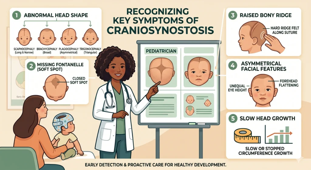
Common Physical Symptoms
An abnormally shaped head serves as the most visible sign. The skull compensates for the fused suture. As a result, the head may look long, narrow, triangular, or flattened on one side.
Other common clinical symptoms include:
- A missing “soft spot” (fontanelle) on the top of the baby’s skull.
- A hard, raised bony ridge running along the fused joint.
- Asymmetrical facial features, such as one eye socket appearing higher or further forward.
- Slow or completely stalled growth in the baby’s overall head circumference.
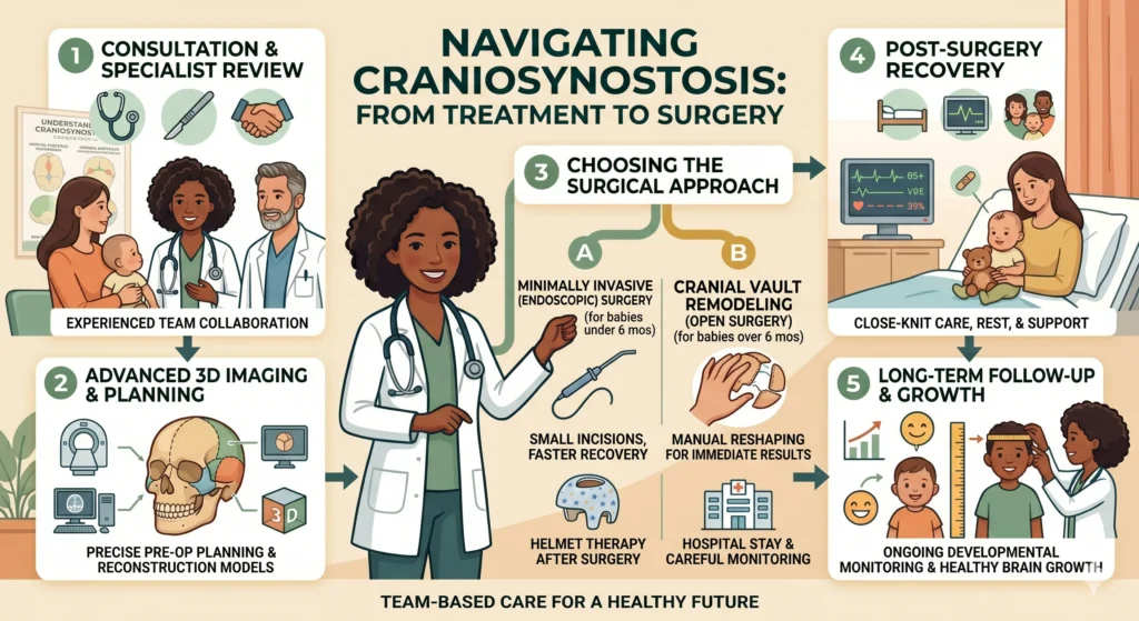
The Diagnostic Process
The diagnostic process usually starts at your pediatrician’s office. A doctor can often identify the condition during a routine physical exam. They feel the baby’s skull for ridges and carefully observe the head shape.
To definitively confirm the diagnosis, the medical team typically orders imaging tests. A CT (Computed Tomography) scan or specialized X-ray provides a highly detailed, 3D look at the bone structure. This allows surgeons to see exactly which sutures fused and plan the surgery accurately.
Differentiating Craniosynostosis from “Flat Head Syndrome”
New parents frequently confuse craniosynostosis with positional plagiocephaly, commonly called “flat head syndrome.”
Both conditions cause an asymmetrical head shape. However, they have entirely different causes and treatments. External pressure causes positional plagiocephaly. Babies develop a flat spot from sleeping or resting in one position for too long.
In positional plagiocephaly, the skull sutures remain completely normal, flexible, and open. The joints function properly, so this condition never requires surgery. Parents can treat it easily through repositioning or increased “tummy time.” In more pronounced cases, doctors prescribe custom molding helmets to gently round out the skull.
Understanding the Causes and Genetic Factors
Parents often wonder if they caused the condition during pregnancy.
Environmental and Genetic Links
In most cases, the exact cause of premature fusion remains unknown. Medical professionals link it to random genetic occurrences and mixed environmental factors.
A smaller percentage involves syndromic cases. These cases link directly to underlying genetic conditions, such as Apert, Crouzon, or Pfeiffer syndrome. Syndromic cases are complex. They often involve multiple fused sutures alongside other physical abnormalities in the hands, feet, or face.
Risks for Future Pregnancies
Does isolated craniosynostosis affect future pregnancies? If your child has non-syndromic craniosynostosis, the chances of a sibling having the condition remain exceedingly low. Doctors consider it a random event.
Conversely, genetic syndromes carry a much higher rate of transmission. We strongly encourage parents facing syndromic cases to consult a genetic counselor. These specialists help families understand the risks for future family planning.
Navigating Treatment and Surgery
When dealing with a fused skull suture, doctors almost always recommend medical intervention.
Why Surgery is Necessary
Surgeons perform operations to achieve two primary goals. First, they relieve any actual or potential pressure on the growing brain. Second, they physically correct the skull deformity to promote normal, symmetrical growth. Medical teams only monitor the most exceptionally mild cases without immediate surgery. Your child’s age and specific fused sutures dictate the chosen surgical path.
Endoscopic Surgery Option
Endoscopic surgery offers a highly effective, minimally invasive approach. Surgeons must perform this procedure while the skull bones remain soft and pliable. Therefore, babies usually undergo this surgery before reaching 3 to 6 months of age.
The surgeon uses an endoscope (a small tube with a camera) to make tiny incisions and carefully open the fused joint. The brain’s rapid growth naturally pushes the skull into shape afterward. Because of this natural reshaping process, babies almost always wear a custom cranial molding helmet for several months post-surgery.
Cranial Vault Remodeling (CVR)
Cranial Vault Remodeling represents the traditional, open-surgery approach. Doctors generally perform CVR when babies grow older and develop thicker skull bones, usually between 6 and 12 months of age.
A multidisciplinary team makes an incision to access the skull. Usually, a pediatric neurosurgeon and a craniofacial plastic surgeon work together. They manually remove, reshape, and replace the affected bone. This immediate reconstruction corrects the head shape and creates vital space for the brain. Children undergoing CVR typically do not need post-operative helmets.
Success Rates and Recovery
Infant surgery feels deeply daunting for any family. However, craniosynostosis surgeries boast exceptionally high success rates.
Surgical complications rarely occur in healthy children. The medical team focuses heavily on managing blood loss during the operation. Specialized pediatric anesthesiologists monitor the child strictly from start to finish.
Partner with an experienced, dedicated pediatric craniofacial team to ensure excellent outcomes. Following the procedure, most children achieve fantastic cosmetic results. They experience healthy, unrestricted brain growth and move forward to live vibrant, unhindered lives.
Let Humane Medical Assistance Bridge the Gap
When a doctor delivers a severe pediatric diagnosis, panic instantly sets in. The prospect of organizing international medical travel, securing visas, and finding the right surgeon while caring for a critically ill infant feels like an impossible, insurmountable mountain.
You do not have to climb that mountain alone.
At Humane Medical Assistance, we serve as your dedicated, deeply compassionate healthcare facilitation partner. We understand the specific cultural and logistical needs of our international patients, and we expertly remove every single barrier between your child and the life-saving care they need.
How We Facilitate Your Child’s Journey to Health:
- Elite Surgeon Selection: We bypass the confusion and connect your family directly with India’s absolute best, proven pediatric neurosurgeons and specialized craniofacial teams.
- Rapid Medical Visas & Logistics: When a child’s brain is under pressure, every day counts. We expedite the complex medical visa process and flawlessly coordinate all airport transfers and ground logistics.
- Safe, Comfortable Accommodations: We secure highly sanitary, comfortable, and private local housing specifically suited for families recovering from major pediatric procedures.
- Transparent, Negotiated Financials: We aggressively advocate on your behalf to secure highly transparent, cost-effective treatment packages, ensuring no hidden fees compound your stress.
- Relentless Patient Advocacy: Our dedicated patient coordinators remain by your side in India 24/7. We manage the hospital bureaucracy so you can focus entirely on holding and comforting your child.
The story of the Tanzanian infant in New Delhi proves that congenital defects and rare diseases do not have the final word. Advanced medical science, flawless surgical execution, and dedicated international coordination can rewrite a child’s future.
Do not let fear or geography delay your child’s treatment. Contact Humane Medical Assistance today. Let us expertly facilitate your journey across borders, guiding your family toward world-class pediatric care and a long, healthy life for your child.